Mosaic Embryo Transfer in Assisted Reproductive Technology (A.R.T)
Mosaicism was initially identified in the field of preimplantation genetic testing for aneuploidy (
PGT-A) approximately 25 years ago. It was initially attributed to an inadequate sample size of trophectoderm (TE) cells. However, technological advancements, particularly the advent of next-generation sequencing (
NGS), have substantially improved our ability to detect and quantify mosaicism. Recently, some experts have suggested that the term 'intermediate copy number' for individual chromosomes is a more precise description than 'mosaicism.'
Mosaic embryos have shown the potential to successfully implant and develop into genetically healthy babies. In a notable study by
Greco et al. in 2015, 18 women who underwent transfers of mosaic embryos gave birth to six healthy newborns with a normal chromosomal makeup. In a recent prospective study, the authors further confirmed the viability of
mosaic embryos by revealing that they had comparable implantation rates (55% vs. 55.8%, p=0.86) and live birth rates (43.4% vs. 42.9%, p=0.82) when compared to euploid embryos. These findings also underscore that mosaic embryos exhibit equivalent developmental potential to their euploid counterparts.
The debate regarding the
transfer of mosaic embryos continues. In 2019, objections were raised by the International Do No Harm Group in in vitro fertilization (
IVF) against the guideline for mosaic embryo transfer issued by the Preimplantation Genetic Diagnosis International Society (
PGDIS). They argued that the interpretation of mosaicism in preimplantation genetic testing for aneuploidy (PGT-A) might be potentially misleading. However, a recent prospective non-selection study presented a different perspective, revealing a remarkably low risk of clinical error (0%–2%) in diagnosing uniform aneuploidy using NGS-based PGT-A. This finding suggests that PGT-A holds significant predictive power.
Definition and types of mosaicism
Mosaicism refers to the presence of multiple genetically distinct cell populations within a single zygote. These mosaic cell populations are believed to result from mitotic errors that occur after the zygote's formation. In the context of Preimplantation Genetic Testing for Aneuploidy (
PGT-A), mosaicism is defined as a mixture of DNA content, with embryos having 20% to 80% aneuploid DNA (an abnormal number of chromosomes) categorized as mosaic. Embryos with less than 20% aneuploid DNA are considered euploid (with the correct number of chromosomes), while those with more than 80% aneuploid DNA are categorized as aneuploid.
The occurrence of mosaic embryos has been reported at approximately 5%, although some studies have found higher rates, ranging from 20% to 30%, when using PGT-A. Mosaicism is more commonly observed in
cleavage-stage embryos (30% to 70%) compared to
blastocyst-stage embryos (5% to 15%).
Mosaicism in the blastocyst stage can be categorized into four types based on cell lineage and the timing of mitotic errors. A blastocyst is termed 'total mosaic' when both the inner cell mass (ICM) and trophectoderm (TE) contain a mix of aneuploid and euploid cells. If only the ICM has the mosaic population, the embryo is referred to as 'ICM mosaic.' Conversely, if only the TE displays mosaicism, the embryo is labeled as 'TE mosaic.' Lastly, when all ICM cells are aneuploid while all TE cells are euploid (or vice versa), the embryo falls into the category of 'ICM/TE mosaic.
Factors Influencing the Detection of Mosaicism
Mosaicism doesn't always exhibit a direct association with maternal age. In fact, some researchers have suggested a slight increase in mosaicism among younger patients when compared to women aged 37 or older. Furthermore, when examining cases of low-degree mosaicism and segmental aneuploidies, the incidence of mosaicism showed a negative correlation with maternal age.
In contrast to the influence of maternal age, a positive link was observed between ovarian response to stimulation and the occurrence of segmental aneuploidy.
A notable prevalence of mosaic embryos has been detected in couples with low sperm concentrations. Among men with oligozoospermia and azoospermia, the occurrence of mosaic and chaotic aneuploidy in blastomeres has been reported to range from 35% to 68%. In Preimplantation Genetic Testing for Aneuploidy (PGT-A) cycles involving cases of
male infertility, a higher proportion of mosaic embryos is observed in comparison to patients with normal sperm parameters. It's worth noting that the severity of male infertility is positively correlated with the rates of mosaicism, with the highest rates observed in cases of severe male infertility.
Several technical laboratory factors have the potential to influence the quality of biopsies, and consequently, they may impact the presence of mosaicism within the trophectoderm (TE). Differences in platform specificity and sensitivity, DNA amplification protocols, and the established threshold settings for interpretation can contribute to variations in mosaicism rates and the quantity of euploid embryos suitable for transfer. Aspects related to the biopsy procedure itself, including loading conditions and the number of cells biopsied, are additional factors that can influence outcomes. Factors such as the method of fertilization and the laboratory environment, including variables like oxygen concentration, pH, osmolality within the embryo culture medium, and temperature, have also been associated with an increased incidence of mosaicism.
Handling: Mosaic Embryo Transfer
The degree of chromosomal mosaicism is categorized into low-level mosaicism when abnormal cells range from 30% to 50% and
high-level mosaicism when abnormal cells constitute 50% to 70%, as defined by the NGS validation algorithm.
In general, embryos with
low-level mosaicism are more likely to develop into healthy babies compared to high-level mosaic embryos. High-level mosaic embryos tend to increase the risk of miscarriage. A recent prospective study demonstrated that embryos with more than 50% mosaicism had significantly lower implantation rates (24.4% vs. 54.6%; p<0.002), clinical pregnancy rates (15.2% vs. 46.4%; p<0.001), and live birth rates (15.2% vs. 46.6%; p<0.001) compared to euploid embryos in the NGS profile.
Capalbo et al. found that low-level (20%–30%) or moderate-degree (30%–50%) mosaic embryo transfers resulted in similar clinical and neonatal outcomes in a prospective double-blinded non-selection trial.
- Specific chromosomes involved.
The clinical impact of mosaicism can significantly vary depending on the specific chromosomes involved. Autosomal chromosomes have been ranked based on their risk of causing placental insufficiency, intrauterine growth restriction, and uniparental disomy (UPD). Mosaic trisomy of
chromosome 16 is frequently encountered in preimplantation embryos and is associated with a high risk of abnormal perinatal outcomes, including intrauterine growth restriction, preterm birth, and hypertensive disorders. Chromosomes X, 21, and 22 have been reported to be susceptible to whole
chromosome errors. Chromosomes 2, 6, 7, 11, 14, 15, 16, and 20 are known to be associated with UPD. Mosaic trisomies of chromosomes 1, 3, 10, 12, and 19 were given the highest priority for
embryo transfer due to their low risk of causing harmful outcomes. Conversely, mosaic trisomies of chromosomes 13, 16, 18, 21, 45, and monosomy X were associated with a high risk of nonviable births and should generally be avoided. However, it's important to note that assessing the degree of mosaicism in preimplantation embryos based solely on molecular and cytogenetic results can be challenging.
Conclusion
While the interest in mosaic embryo transfers is increasing, the ongoing debate about whether such transfers are appropriate continues. In clinical practice, it is crucial to identify viable and suitable mosaic subgroups for transfer. Equally important is the need to inform patients that data on postnatal and neonatal outcomes after mosaic embryo transfers remain limited, and clinical results have shown variation.
Esco Medical offers a comprehensive range of advanced tools, which include
ART workstations,
benchtop incubators, and
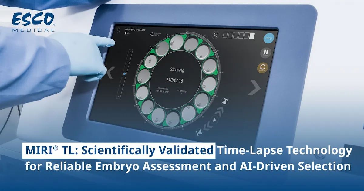
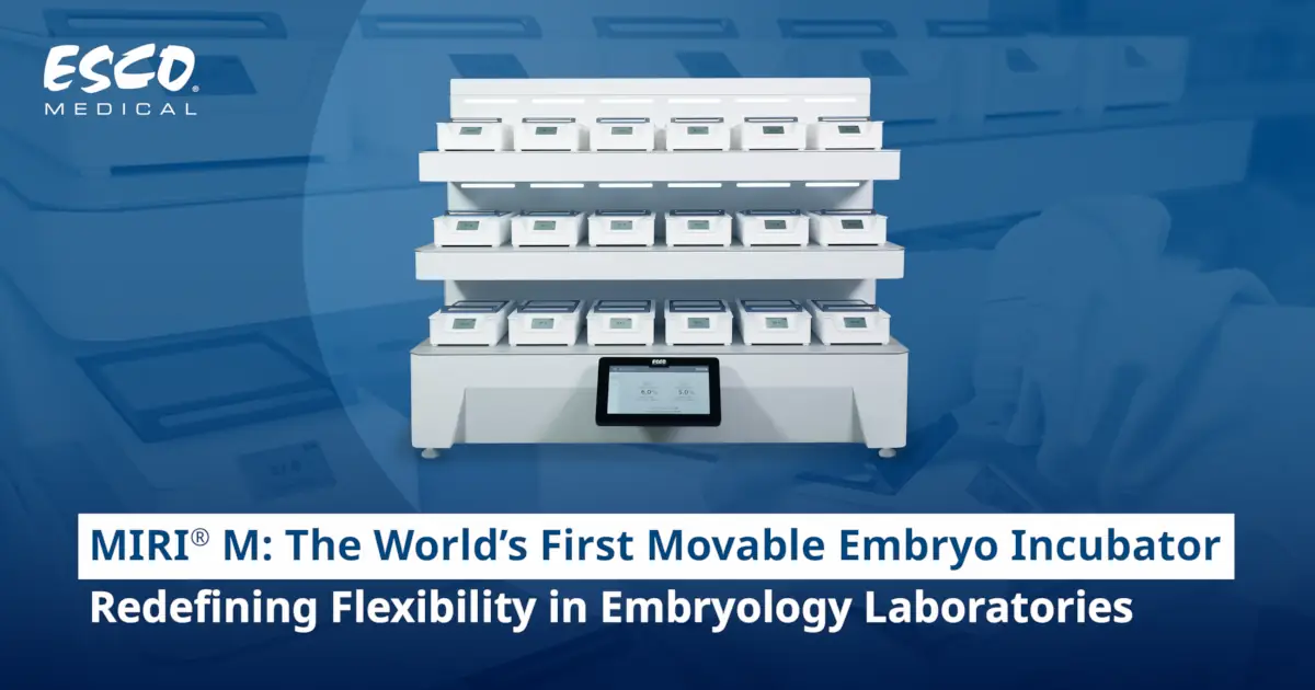
time-lapse systems. These tools are meticulously designed to create the ideal environment for preimplantation embryo development. Esco Medical's equipment plays a pivotal role in maintaining the culture conditions necessary for fostering the healthy development of embryos and reducing the risk of mosaicism.
